


Sabine Steidl, RGZM/Burkhard Schillinger, MLZ
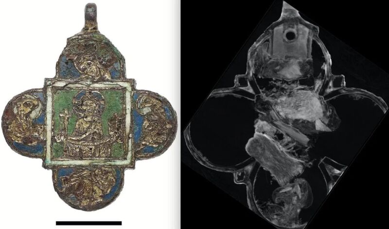
In 2008, archaeologists excavating a medieval dump in Mainz, Germany, found a severely corroded pendant thought to have been made in the late 12th century. However, they were reluctant to open the pendant to see what was inside. Now technology has helped. Researchers at the Technical University of Munich used neutron tomography and other methods to scan the pendant and found that it contained bone fragments. The findings were presented at an interim meeting of the International Council of Museums and Conservation Commission (ICOM-CC) Working Group on Metals.
Neutron tomography works much like X-ray and gamma-ray imaging, except that it uses a neutron beam. One shoots a beam of radiation at the target, some parts interacting with the sample and other parts passing through. The latter collides with the imaging target to create what is called an attenuation pattern, an image of the interior of the sample. Neutron tomography is not as sensitive to material density as X-ray and gamma-ray imaging, and unlike these methods, neutrons interact strongly with very light elements such as hydrogen. So what is easily visible in neutron imaging may be difficult or impossible to see in X-ray imaging (and vice versa).
These techniques are complementary and do not damage or destroy the original object, making them particularly useful for imaging archaeological or paleontological artifacts. For example, in December 2021, the researcher combined his X-ray microtomography (which involves using X-rays to create cross-sections of objects) and neutron tomography to reveal a very large 365-million-year-old ammonite. created a detailed 3D model of him. Jurassic fossils reveal previously unseen internal muscles. Among other findings, they observed paired muscles extending from the ammonite’s body. This suggests that ammonites were likely used to pull their bodies into their shells to avoid predators.
Sabine Steidl, RGZM
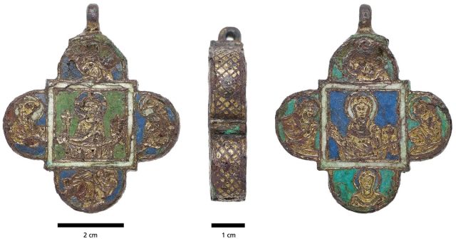
The gold-plated copper pendant in Mainz is only 2.4 inches (6 cm) high and wide and shaped like a quatrefoil (a common form in traditional Christian symbolism). The front and back are enamelled using a technique called champlevé. Champlevé is the carving or etching of valleys in a metal surface and filling them with porcelain enamel. The uncovered parts are plated with gold, a common practice in the Middle Ages. One side depicts Jesus and the four rounded edges depict his four evangelists. The other side shows Mary surrounded by four female saints.
The team first analyzed the surface using a combination of micro X-ray fluorescence and Raman spectroscopy to identify all the elements present. Infrared spectroscopy also revealed a small sample of beeswax. But Matthias Heinzel, restorer of his Leibniz-Zentrumfür Archäologie (LEIZA), part of the Technical University of Munich, said: The mechanism has been severely damaged by centuries of corrosion, and opening it would mean irreparable destruction. “
Using neutron imaging, they revealed five small reliquary boxes of silk and linen in which pendants were preserved and which held bone fragments.Heinsel othersIndividual elements of the samples were identified by triggering them with a gamma-ray technique called prompted gamma activation analysis (PGAA). “I don’t know if these bone fragments are from a saint, and if so, I don’t know which one,” Heintzel said. “Normally, relic packages would include a strip of parchment indicating the saint’s name. But in this case, unfortunately, we can’t see it.”
The now fully restored pendant is now on display at the Mainz State Museum.