










UNSW researchers have unveiled a prototype device that 3D-prints living cells directly into internal organs and could potentially be used as an all-in-one endoscopic surgical tool.
Engineers at UNSW Sydney have developed a small, flexible, soft robotic arm that can be used to 3D print biomaterials directly into organs inside the human body.
3D bioprinting is the process of manufacturing biomedical components from so-called bioinks to build tissue-like structures.
Bioprinting is mainly used for research purposes such as tissue engineering and for the development of new drugs. Typically, generating cell structures in vitro requires the use of large 3D printing machines.
The new study at the UNSW Medical Robotics Lab is led by Dr Thanh Nho Do and his PhD student Mai Thanh Thai, and includes Science Professor Nigel Lovell, Dr Hoang-Phuong Phan, and Associate Professor Jelena Rnjak-Kovacina. was conducted in collaboration with researchers in It is described in detail in a paper published in Advanced Science.
Their work has led to a small, flexible 3D bioprinter that can be inserted into the body like an endoscope and deliver multi-layered biomaterials directly to internal organs and tissue surfaces.
The proof-of-concept device, known as F3DB, features a highly maneuverable swivel head that “prints” bioink, attached to the end of a long, flexible, snake-like robotic arm, all controlled externally. can.
With further development, the researchers believe, potentially within 5 to 7 years, medical professionals will be able to use the technology to access hard-to-reach areas of the body through small skin incisions or natural openings. It says it may be accessible.
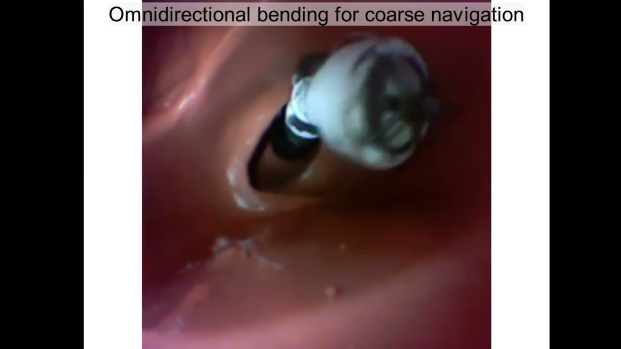
The research team tested the device inside an artificial colon, which allows it to pass through confined spaces, before successfully 3D printing it.
Dr. Daw and his team tested the device in an artificial colon and 3D printed a variety of materials in different shapes onto the surface of pig kidneys.
“Existing 3D bioprinting technologies require biomaterials to be created outside the body, and transplanting them into humans typically requires extensive open-field open surgery, which increases the risk of infection.” , Scientia Senior Lecturer in Biomedical Engineering (GSBmE) and Tyree Foundation Institute of Health Engineering (IHealthE) at UNSW.
“Our flexible 3D bioprinter means we can deliver biomaterials directly to target tissues or organs with a minimally invasive approach.
“This system offers the possibility of accurately reconstructing three-dimensional wounds in the body, such as damage to the stomach wall or damage or disease in the colon.
“Thanks to its flexible body, our prototype can 3D print multi-layered biomaterials of various sizes and shapes through restricted and hard-to-reach areas.
“Our approach overcomes the significant challenges of existing 3D bioprinters, such as surface mismatch between the 3D-printed biomaterial and the target tissue/organ, and structural damage during manual handling, transfer, and transportation processes. It also addresses restrictions.”
Scientia Professor Nigel Lovell, Head of GSBmE and Director of IHealthE, added: Several other proof-of-concept devices have been presented, but are much more rigid and unwieldy for use in complex and confined spaces within the body. ”
The smallest F3DB prototype built by the UNSW team has a diameter similar to that of commercial therapeutic endoscopes (about 11-13 mm) and is small enough to be inserted into the human gastrointestinal tract.
But researchers say they could easily be made even smaller for future medical applications.
soft robotics
This device features a 3-axis print head mounted directly on the tip of a soft robotic arm. Composed of soft artificial muscles and moving in three directions, the print head works much like a traditional desktop 3D printer.
Flexible robotic arms can be hydraulically bent and twisted and can be manufactured in any length you need. Its stiffness can be fine-tuned using different types of elastic tubing and fabrics.
The print nozzle can be programmed to print pre-determined shapes or manually operated when more complex or uncertain bioprinting is required. Additionally, the team utilized a machine learning-based controller that can assist with the printing process.
To further demonstrate the feasibility of this technology, the UNSW team tested the cell viability of living biomaterials after being printed through the system.
In our experiments, we observed that the cells were unaffected by the process and most of the cells were alive after printing. Cells then continued to proliferate for the next seven days, with four times as many cells observed one week after printing.
All-in-one endoscopic surgical instrument
The research team also demonstrated that F3DB could potentially be used as an all-in-one endoscopic surgical tool to perform a variety of functions.
They say this may be of particular importance in surgery to resect certain cancers, particularly colorectal cancer, via a process known as endoscopic submucosal dissection (ESD).
Globally, colorectal cancer is the third leading cause of cancer death, and early resection of colorectal tumors improves patients’ 5-year survival by at least 90%.
The nozzle of the F3DB printhead can be used as a kind of electrocautery to first mark cancerous lesions and then excise them.
You can also run water through a nozzle to wash blood and excess tissue from the area at the same time. Also, 3D printing biomaterials directly while the robotic arm is still in place can promote faster healing.

The research team has demonstrated that F3DB can be used in a variety of ways when developed as an all-in-one endoscopic surgical tool.
The ability to perform such multifunctional procedures has been demonstrated in porcine intestines, researchers say, indicating that F3DB is a promising candidate for future development of an all-in-one endoscopic surgical tool. increase.
“Compared to existing endoscopic surgical tools, the developed F3DB was designed as an all-in-one endoscopic tool, avoiding the use of modifiable tools that are typically associated with longer procedure times and infection risks. I will,” said Mai Thanh Thai.
The next step in the development of this provisionally patented system is in vivo testing in live animals to demonstrate its utility.
The researchers also plan to implement additional features such as an integrated camera and real-time scanning system to reconstruct 3D tomography of moving tissue inside the body.
Original: 3D bioprinting inside the human body could be possible thanks to new soft robots
Than: University of New South Wales